ANATOMY OF TESTIS
- The testis is composed of a multitude of small, intricately coiled tubules known as seminiferous tubules. The aforementioned structures comprise the spermatogenic tissue found within the testis.
- The tubules are lined with cells that undergo spermatogenesis, resulting in the production of spermatozoa that are subsequently released into the tubule lumen.
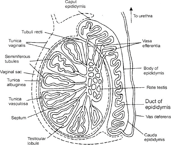
Structure of testis
- Within the context of spermatogenesis, the presence of Sertoli cells, also known as sustentacular or nurse cells, can be observed. These cells play a crucial role in providing nourishment to the developing spermatozoa and regulating the process of spermatogenesis. Additionally, they release inhibin to effectively control the excessive activity of follicle-stimulating hormone (FSH).
- The additional roles performed by Sertoli cells encompass several functions.
- Firstly, they provide nourishment to the maturing spermatozoa.
- Secondly, they absorb the components that are shed during the development of spermatozoa.
- Thirdly, they release anti-Mullerian factor (AMF) in order to inhibit the development of the Mullerian duct or oviduct.
- Lastly, they release androgen binding protein (ABP).
- Groups of polyhedral cells known as Interstitial cells or Leydig cells are situated within the connective tissue surrounding the seminiferous tubules.
- The testicular endocrine tissue is comprised by them. The Leydig cells are responsible for the secretion of testosterone into the bloodstream.
- The seminiferous tubules coalesce to generate multiple linear tubules known as tubuli recti, which subsequently connect to irregular cavities located in the posterior region of the testis.
- The rete testis is composed of a complex network of interconnected channels lined with cuboidal epithelium, exhibiting a highly anastomosing structure.
- Multiple tubules known as vasa efferentia originate from the testis and facilitate the transportation of spermatozoa out of the testis. These vasa efferentia form an intratesticular genital duct system connecting the seminiferous tubules to the vasa efferentia.
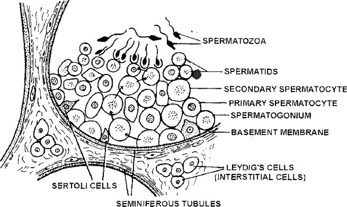
A part of transverse section of mammalian testis
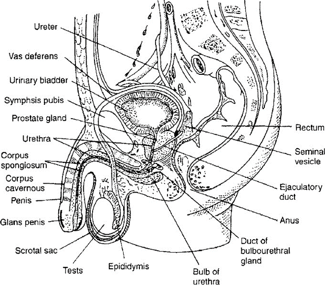
Midline sagittal section through male pelvis
- The extratesticular duct system comprises a series of tubules that facilitate the transportation of sperm from the testes to the external environment.
- The process commences with the vasa efferentia, which originate from each testis and subsequently merge to create a convoluted and folded structure known as the epididymis, situated posteriorly to each testis.
- The epididymis is comprised of three distinct components, namely the Caput, Corpus, and Cauda. The epididymis serves as a temporary storage site for sperm.
- The cauda epididymis gives rise to a partially coiled tube known as the vas deferens, which ascends into the abdomen via the inguinal canal. It traverses over the urinary bladder and merges with the duct from the seminal vesicle located posterior to the urinary bladder, forming an ejaculatory duct.
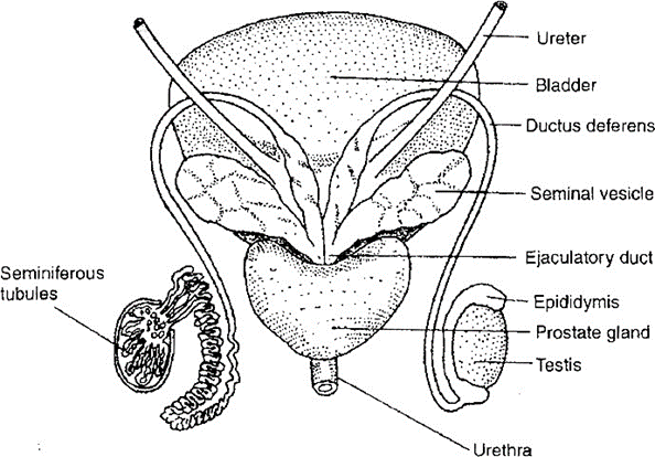
The male genital system seen from behind
- Prior to entering the prostate, the ductus deferens undergoes dilation to form an ampulla. The terminal segment of the ampulla traverses the prostate gland, ultimately connecting with the urethra in close proximity to its point of origin from the urinary bladder.
- The urethra serves as the recipient of the ducts originating from the prostate and Cowper’s glands. It traverses the penis and ultimately terminates at the external opening.